
Every tissue in the human body contains exceptionally small fibers that help coordinate how organs move, function and communicate. Muscle fibers guide physical force, intestinal fibers support the motion of the digestive tract, and brain fibers carry electrical signals that allow different regions to exchange information. Together, these intricate fiber systems help shape the structure of each organ and keep them operating properly.
[…]
Although these microscopic structures play essential roles, they have long been challenging to study. Researchers have struggled to determine how fibers are oriented inside tissues, which has made it difficult to fully understand how they change in health and disease.
A Simple Method for Revealing Hidden Microstructure
A research team led by Marios Georgiadis, PhD, instructor of neuroimaging, has now introduced an approach that makes these hard-to-see fiber patterns visible with exceptional clarity and at a relatively low cost.
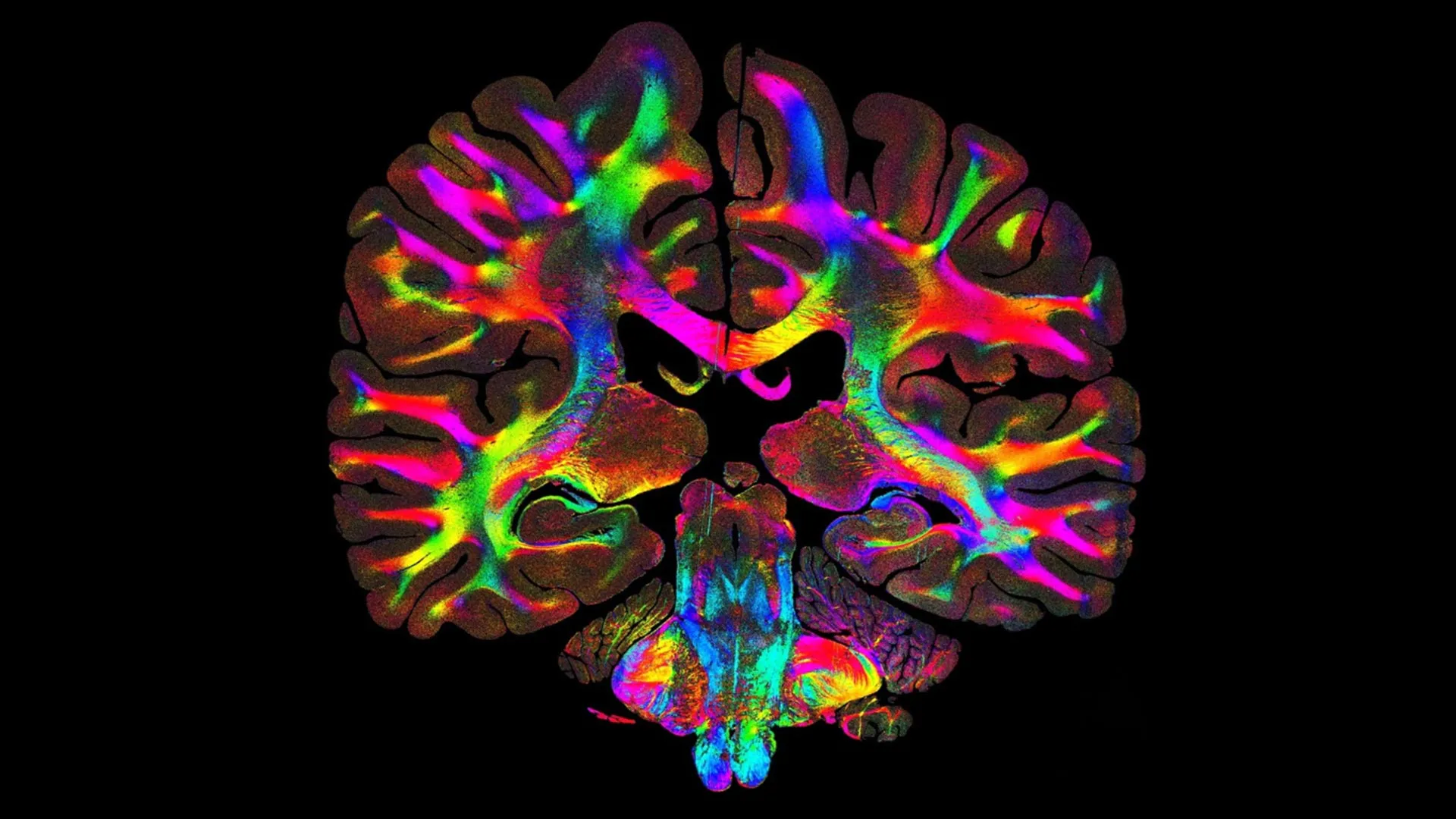
Their technique, described in Nature Communications, is known as computational scattered light imaging (ComSLI). It can reveal the orientation and organization of tissue fibers at micrometer resolution on virtually any histology slide, regardless of how it was stained or preserved — even if the slide is many decades old.
[…]
ComSLI relies on a basic physical principle: when light encounters microscopic structures, it scatters in different directions based on their orientation. By rotating the light source and recording how the scattering signal changes, researchers can reconstruct the direction of the fibers within each pixel of an image.
The method requires only a rotating LED light and a microscope camera, making the setup accessible compared with other forms of advanced microscopy. After the images are collected, software analyzes delicate patterns in the scattered light to generate color-coded maps of fiber orientation and density, known as microstructure-informed fiber orientation distributions.
ComSLI is not limited by sample preparation. It works with formalin-fixed, paraffin-embedded sections (a standard in hospitals and pathology labs) as well as fresh-frozen, stained or unstained slides.
[…]
“This is a tool that any lab can use,” Zeineh said. “You don’t need specialized preparation or expensive equipment. What excites me most is that this approach opens the door for anyone, from small research labs to pathology labs, to uncover new insights from slides they already have.”
[…]
To test the limits of the method, the researchers analyzed a brain section prepared in 1904. Even in this century-old sample, ComSLI identified intricate fiber patterns, allowing scientists to study historical specimens and explore how structural features evolve across generations of disease.
Applications Beyond the Brain
Although first designed for brain research, ComSLI also works well in other tissues. The team used it to study muscle, bone and vascular samples, each revealing unique fiber arrangements tied to their biological functions.
In tongue muscle, the method highlighted layered fiber orientations linked to movement and flexibility. In bone, it captured collagen fibers that align with mechanical stress. In arteries, it showed alternating collagen and elastin layers that support both strength and elasticity.
This ability to map fiber orientation across species, organs and archival specimens could significantly change how scientists investigate structure and function.
[…]
Story Source:
Materials provided by Stanford Medicine. Note: Content may be edited for style and length.
Journal Reference:
- Marios Georgiadis, Franca auf der Heiden, Hamed Abbasi, Loes Ettema, Jeffrey Nirschl, Hossein Moein Taghavi, Moe Wakatsuki, Andy Liu, William Hai Dang Ho, Mackenzie Carlson, Michail Doukas, Sjors A. Koppes, Stijn Keereweer, Raymond A. Sobel, Kawin Setsompop, Congyu Liao, Katrin Amunts, Markus Axer, Michael Zeineh, Miriam Menzel. Micron-resolution fiber mapping in histology independent of sample preparation. Nature Communications, 2025; 16 (1) DOI: 10.1038/s41467-025-64896-9
Source: Simple light trick reveals hidden brain pathways in microscopic detail | ScienceDaily

Robin Edgar
Organisational Structures | Technology and Science | Military, IT and Lifestyle consultancy | Social, Broadcast & Cross Media | Flying aircraft