A common supplement could supercharge cancer treatments

In a new study, researchers from the University of Chicago discovered that zeaxanthin, a plant-derived carotenoid best known for protecting vision, may also act as an immune-boosting compound by strengthening the cancer-fighting activity of immune cells. The findings, which were published in Cell Reports Medicine, highlight the potential of zeaxanthin as a widely available supplement to improve the effectiveness of cancer immunotherapies.

“We were surprised to find that zeaxanthin, already known for its role in eye health, has a completely new function in boosting anti-tumor immunity,” said Jing Chen, PhD, Janet Davison Rowley Distinguished Service Professor of Medicine and senior author of the study. “Our study show that a simple dietary nutrient could complement and strengthen advanced cancer treatments like immunotherapy.”

How does this nutrient work?

The study builds on years of work by Chen’s lab to better understand how nutrients influence the immune system. By screening a large blood nutrient library, the team identified zeaxanthin as a compound that directly enhances the activity of CD8+ T cells, a crucial type of immune cell that kills tumor cells. These cells rely on a molecular structure called the T-cell receptor (TCR) to recognize and destroy abnormal cells.

The researchers found that zeaxanthin stabilizes and strengthens the formation of TCR complex on CD8+ T cells upon interacting with the cancer cells. This, in turn, triggers more robust intracellular signaling that boosts T-cell activation, cytokine production, and tumor-killing capacity.

Zeaxanthin improves immunotherapy effects

In mouse models, dietary supplementation with zeaxanthin slowed tumor growth. Importantly, when combined with immune checkpoint inhibitors – a type of immunotherapy that has transformed cancer treatment in recent years – zeaxanthin significantly enhanced anti-tumor effects compared to immunotherapy alone.

To extend the findings, the researchers tested human T cells engineered to recognize specific tumor antigens and found that zeaxanthin treatment improved these cells’ ability to kill melanoma, multiple myeloma, and glioblastoma cells in laboratory experiments.

[…]

`

Zeaxanthin is sold as an over-the-counter supplement for eye health, and is naturally found in vegetables like orange peppers, spinach, and kale. It’s inexpensive, widely available, well-tolerated and, most importantly, its safety profile is known – which means it can be safely tested as an adjunct to cancer therapies.

The study also reinforces the importance of a balanced diet. In their previous research, Chen’s group discovered that trans-vaccenic acid (TVA), a fatty acid derived from dairy and meat, also boosts T-cell activity – but through a different mechanism. Together, the findings suggest that nutrients from both plant and animal sources may provide complementary benefits to immune health.

Clinical applications of zeaxanthin

Although the results are promising, the researchers emphasize that the work is still at an early stage. Most of the findings come from laboratory experiments and animal studies. Thus, clinical trials will be needed to determine whether zeaxanthin supplements can improve outcomes for cancer patients.

[…]

Source: A common supplement could supercharge cancer treatments | ScienceDaily

Scientists develop ‘glue gun’ that 3D prints bone grafts directly onto fractures

Researchers have modified a standard glue gun to 3D print a bone-like material directly onto fractures, paving the way for its use in operating rooms.

The device, which has so far been tested in rabbits, would be particularly useful for fixing irregularly shaped fractures during surgery, the researchers say.

[…]

Typically, large fractures or other defects in bones require bone grafting and the use of a metal fastener, such as a pin or plate, to support the broken bone as it heals. But because the shape of these implants is not specific to a given patient’s fracture, this can result in poor alignment and compromised stability of the bone.

Previous studies have shown that 3D printing bespoke bone grafts for individual patients can be done, but these required considerable time and effort to make, preventing them from being used on the fly during surgery.

Now, in a study published Friday (Sept. 5) in the journal Device, Lee and colleagues detailed their new method of 3D printing a bone substitute directly onto a fracture using a glue gun.

Source: Scientists develop ‘glue gun’ that 3D prints bone grafts directly onto fractures | Live Science

New drug could be first to stop deadly fatty liver disease

Researchers at University of California San Diego School of Medicine have identified a new investigational drug that shows promise in treating metabolic dysfunction-associated steatohepatitis (MASH), a serious form of fatty liver disease linked to obesity and type 2 diabetes that can lead to cirrhosis, liver failure, and even liver cancer.

The study, published in the August 23, 2025 online edition of The Lancet, found that the medication, ION224, targets a liver enzyme called DGAT2, which plays a key role in how the liver produces and stores fat. By blocking this enzyme, the drug helps reduce fat buildup and inflammation, two major drivers of liver damage in MASH.

“This study marks a pivotal advance in the fight against MASH,” said Rohit Loomba, MD, principal investigator of the study and chief of the Division of Gastroenterology and Hepatology at UC San Diego School of Medicine. “By blocking DGAT2, we’re interrupting the disease process at its root cause, stopping fat accumulation and inflammation right in the liver.”

The multicenter, Phase IIb clinical trial involved 160 adults with MASH and early to moderate fibrosis across the United States. Participants received monthly injections of the drug at different doses or a placebo over the course of one year. At the highest dose, 60% showed notable improvements in their liver health compared to the placebo group. These benefits occurred regardless of weight change, suggesting the drug could be used alongside other therapies. The medicine showed no serious side effects linked to the treatment.

MASH, formally known as nonalcoholic steatohepatitis (NASH), affects people with metabolic conditions like obesity and type 2 diabetes. It is often called a “silent” disease because it can progress for years without symptoms.

More than 100 million people have some form of fatty liver disease in the U.S. and as many as 1 in 4 adults worldwide may be affected, according to the Centers for Disease Control and Prevention. If left untreated, MASH can progress to liver failure and often may require a transplant.

“This is the first drug of its kind to show real biological impact in MASH,” Loomba said. “If these findings are confirmed in Phase III trials, we may finally be able to offer patients a targeted therapy that halts and potentially reverses liver damage before it progresses to life-threatening stages.”

Source: New drug could be first to stop deadly fatty liver disease | ScienceDaily

Scientists figure out why the flu is deadly for older patients

Scientists have discovered why older people are more likely to suffer severely from the flu, and can now use their findings to address this risk.

In a new study, which is published in PNAS, experts discovered that older people produce a glycosylated protein called apoplipoprotein D (ApoD), which is involved in lipid metabolism and inflammation, at much higher levels than in younger people. This has the effect of reducing the patient’s ability to resist virus infection, resulting in a more serious disease outcome.

The team established that highly elevated ApoD production with age in the lung drives extensive tissue damage during infection to reduce the protective antiviral type I interferon response.

[…]

They identified ApoD as an age-related cell factor that impairs the activation of the immune system’s antiviral response to influenza virus infection by causing extensive breakdown of mitochondria (mitophagy) resulting in greater production of virus and lung damage during infection. Mitochondria are essential for cellular production of energy and for induction of protective interferons.

ApoD is therefore a target for therapeutic intervention to protect against severe influenza virus infection in the elderly which would have a major impact on reducing morbidity and mortality in the aging population.

[…]

Source: Why the flu turns deadly for older adults, and how scientists found the cause | ScienceDaily

Rats walk again after spinal cord repair with 3D printing

[…] a groundbreaking process that combines 3D printing, stem cell biology, and lab-grown tissues for spinal cord injury recovery.

[…]

A major challenge is the death of nerve cells and the inability for nerve fibers to regrow across the injury site. This new research tackles this problem head-on.

The method involves creating a unique 3D-printed framework for lab-grown organs, called an organoid scaffold, with microscopic channels. These channels are then populated with regionally specific spinal neural progenitor cells (sNPCs), which are cells derived from human adult stem cells that have the capacity to divide and differentiate into specific types of mature cells.

“We use the 3D printed channels of the scaffold to direct the growth of the stem cells, which ensures the new nerve fibers grow in the desired way,

[…]

In their study, the researchers transplanted these scaffolds into rats with spinal cords that were completely severed. The cells successfully differentiated into neurons and extended their nerve fibers in both directions — rostral (toward the head) and caudal (toward the tail) — to form new connections with the host’s existing nerve circuits.

The new nerve cells integrated seamlessly into the host spinal cord tissue over time, leading to significant functional recovery in the rats.

[…]

Source: Rats walk again after breakthrough spinal cord repair with 3D printing | ScienceDaily

Forget LASIK: Safer, cheaper vision correction could be coming soon

[…] In the body, the shapes of many collagen-containing tissues, including corneas, are held in place by attractions of oppositely charged components. These tissues contain a lot of water, so applying an electric potential to them lowers the tissue’s pH, making it more acidic. By altering the pH, the rigid attractions within the tissue are loosened and make the shape malleable. When the original pH is restored, the tissue is locked into the new shape.

Previously, the researchers used EMR to reshape cartilage-rich rabbit ears, as well as alter scars and skin in pigs. But one collagen-rich tissue that they were eager to explore was the cornea.

In this work, the team constructed specialized, platinum “contact lenses” that provided a template for the corrected shape of the cornea, then placed each over a rabbit eyeball in a saline solution meant to mimic natural tears. The platinum lens acted as an electrode to generate a precise pH change when the researchers applied a small electric potential to the lens. After about a minute, the cornea’s curvature conformed to the shape of the lens — about the same amount of time LASIK takes, but with fewer steps, less expensive equipment and no incisions.

They repeated this setup on 12 separate rabbit eyeballs, 10 of which were treated as if they had myopia, or nearsightedness. In all the “myopic” eyeballs, the treatment dialed in the targeted focusing power of the eye, which would correspond to improved vision. The cells in the eyeball survived the treatment, because the researchers carefully controlled the pH gradient. Additionally, in other experiments, the team demonstrated that their technique might be able to reverse some chemical-caused cloudiness to the cornea — a condition that is currently only treatable through a complete corneal transplant.

Though this initial work is promising, the researchers emphasize that it is in its very early stages. Next up is what Wong describes as, “the long march through animal studies that are detailed and precise,” including tests on a living rabbit rather than just its eyeball. They also plan to determine the types of vision correction possible with EMR, such as near- and far-sightedness and astigmatism. Though the next steps are planned, uncertainties in the team’s scientific funding have put them on hold.

[…]

Source: Forget LASIK: Safer, cheaper vision correction could be coming soon | ScienceDaily

A new mRNA cancer vaccine just wiped out tumors in mice

An experimental mRNA vaccine boosted the tumor-fighting effects of immunotherapy in a mouse-model study, bringing researchers one step closer to their goal of developing a universal vaccine to “wake up” the immune system against cancer.

Published recently in Nature Biomedical Engineering, the University of Florida study showed that like a one-two punch, pairing the test vaccine with common anticancer drugs called immune checkpoint inhibitors triggered a strong antitumor response.

A surprising element, researchers said, was that they achieved the promising results not by attacking a specific target protein expressed in the tumor, but by simply revving up the immune system — spurring it to respond as if fighting a virus. They did this by stimulating the expression of a protein called PD-L1 inside of tumors, making them more receptive to treatment. The research was supported by multiple federal agencies and foundations, including the National Institutes of Health.

[…]

“This paper describes a very unexpected and exciting observation: that even a vaccine not specific to any particular tumor or virus — so long as it is an mRNA vaccine — could lead to tumor-specific effects,” said Sayour, principal investigator at the RNA Engineering Laboratory within UF’s Preston A. Wells Jr. Center for Brain Tumor Therapy.

“This finding is a proof of concept that these vaccines potentially could be commercialized as universal cancer vaccines to sensitize the immune system against a patient’s individual tumor,” said Sayour, a McKnight Brain Institute investigator and co-leader of a program in immuno-oncology and microbiome research.

Until now, there have been two main ideas in cancer-vaccine development: To find a specific target expressed in many people with cancer, or to tailor a vaccine that is specific to targets expressed within a patient’s own cancer.

“This study suggests a third emerging paradigm,” said Duane Mitchell, M.D., Ph.D., a co-author of the paper. “What we found is by using a vaccine designed not to target cancer specifically but rather to stimulate a strong immunologic response, we could elicit a very strong anticancer reaction. And so this has significant potential to be broadly used across cancer patients — even possibly leading us to an off-the-shelf cancer vaccine.”

[…]

Source: A new cancer vaccine just wiped out tumors in mice | ScienceDaily

Trojan horse bacteria sneak cancer-killing viruses into tumors

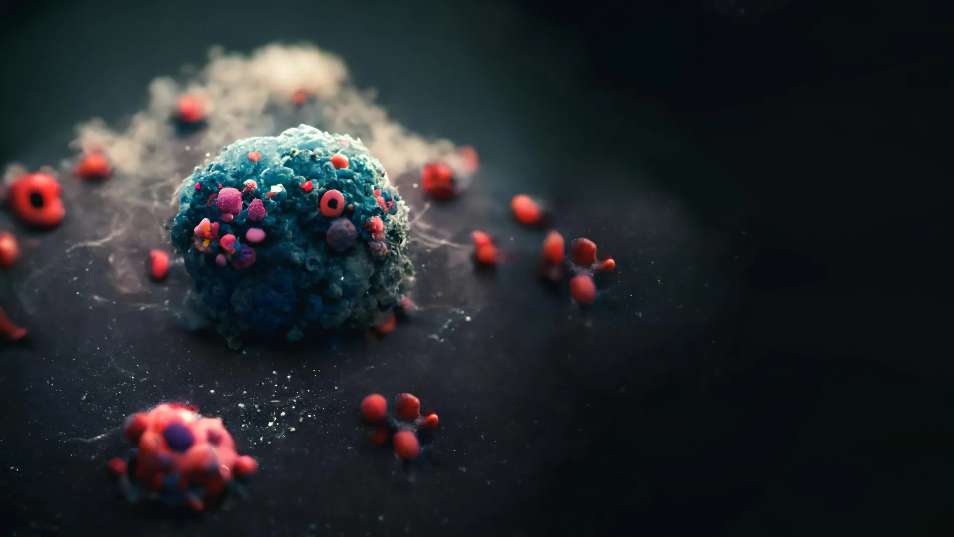
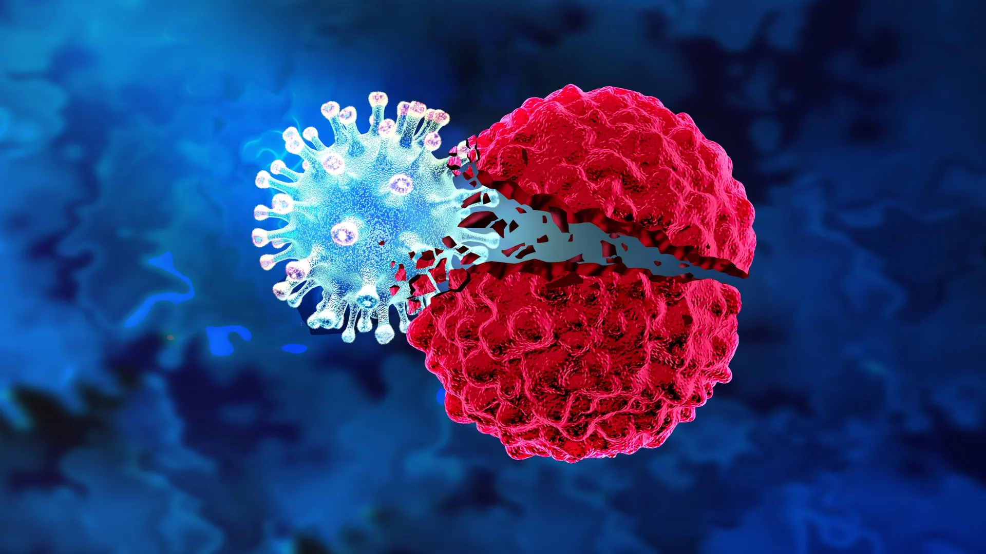
Researchers at Columbia Engineering have built a cancer therapy that makes bacteria and viruses work as a team. In a study published recently in Nature Biomedical Engineering, the Synthetic Biological Systems Lab shows how their system hides a virus inside a tumor-seeking bacterium, smuggles it past the immune system, and unleashes it inside cancerous tumors.

The new platform combines the bacteria’s tendency to find and attack tumors with the virus’s natural preference for infecting and killing cancerous cells. Tal Danino, an associate professor of biomedical engineering at Columbia Engineering, led the team’s effort to create the system, which is called CAPPSID (short for Coordinated Activity of Prokaryote and Picornavirus for Safe Intracellular Delivery). Charles M. Rice, an expert in virology at The Rockefeller University, collaborated with the Columbia team.

“We aimed to enhance bacterial cancer therapy by enabling the bacteria to deliver and activate a therapeutic virus directly inside tumor cells, while engineering safeguards to limit viral spread outside the tumor,” says co-lead author Jonathan Pabón, an MD/PhD candidate at Columbia.

The researchers believe that this technology — validated in mice — represents the first example of directly engineered cooperation between bacteria and cancer-targeting viruses.

The approach combines the bacteria’s instinct for homing in on tumors with a virus’s knack for infecting and killing cancer cells. “By bridging bacterial engineering with synthetic virology, our goal is to open a path toward multi-organism therapies that can accomplish far more than any single microbe could achieve alone,” says Zakary S. Singer, a co-lead author and former postdoctoral researcher in Tal Danino’s lab.

“This is probably our most technically advanced and novel platform to date,” says Danino, who is also affiliated with the Herbert Irving Comprehensive Cancer Center at Columbia University Irving Medical Center and Columbia’s Data Science Institute.

Sneaking past the immune system

One of the biggest hurdles in oncolytic virus therapy is the body’s own defense system. If a patient has antibodies against the virus — from a prior infection or vaccination — those antibodies can neutralize it before it reaches a tumor. The Columbia team sidestepped that problem by tucking the virus inside tumor-seeking bacteria.

“The bacteria act as an invisibility cloak, hiding the virus from circulating antibodies, and ferrying the virus to where it is needed,” Singer says.

Pabón says this strategy is especially important for viruses that people are already exposed to in daily life.

“Our system demonstrates that bacteria can potentially be used to launch an oncolytic virus to treat solid tumors in patients who have developed immunity to these viruses,” he says.

Targeting the tumor

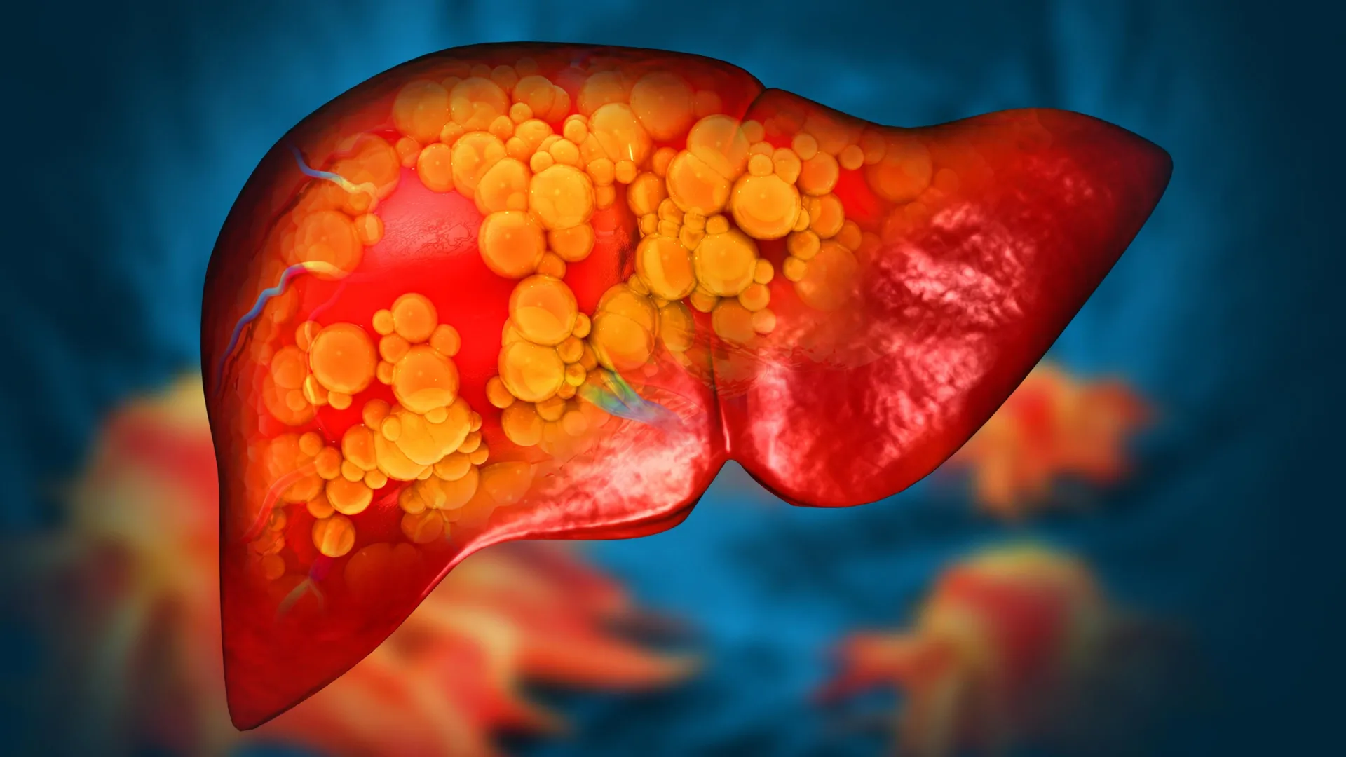
The system’s bacterial half is Salmonella typhimurium, a species that naturally migrates to the low-oxygen, nutrient-rich environment inside tumors. Once there, the bacteria invade cancer cells and release the virus directly into the tumor’s interior.

“We programmed the bacteria to act as a Trojan horse by shuttling the viral RNA into tumors and then lyse themselves directly inside of cancer cells to release the viral genome, which could then spread between cancer cells,” Singer says.

By exploiting the bacteria’s tumor-homing instincts and the virus’s ability to replicate inside cancer cells, the researchers created a delivery system that can penetrate the tumor and spread throughout it — a challenge that has limited both bacteria- and virus-only approaches.

Safeguarding against runaway infections

A key concern with any live virus therapy is controlling its spread beyond the tumor. The team’s system solved that problem with a molecular trick: making sure the virus couldn’t spread without a molecule it can only get from the bacteria. Since the bacteria stay put in the tumor, this vital component (called a protease) isn’t available anywhere else in the body.

“Spreadable viral particles could only form in the vicinity of bacteria, which are needed to provide special machinery essential for viral maturation in the engineered virus, providing a synthetic dependence between microbes,” Singer says. That safeguard adds a second layer of control: even if the virus escapes the tumor, it won’t spread in healthy tissue.

“It is systems like these — specifically oriented towards enhancing the safety of these living therapies — that will be essential for translating these advances into the clinic,” Singer says.

Further research and clinical applications

This publication marks a significant step toward making this type of bacteria-virus system available for future clinical applications.

“As a physician-scientist, my goal is to bring living medicines into the clinic,” Pabón says. “Efforts toward clinical translation are currently underway to translate our technology out of the lab.”

Danino, Rice, Singer, and Pabón have filed a patent application (WO2024254419A2) with the U.S. Patent and Trademark Office related to this work.

Looking ahead, the team is testing the approach in a wider range of cancers, using different tumor types, mouse models, viruses, and payloads, with an eye to developing a “toolkit” of viral therapies that can sense and respond to specific conditions inside a cell. They are also evaluating how this system can be combined with strains of bacteria that have already demonstrated safety in clinical trials.


Story Source:

Materials provided by Columbia University School of Engineering and Applied Science. Note: Content may be edited for style and length.


Journal Reference:

  1. Zakary S. Singer, Jonathan Pabón, Hsinyen Huang, William Sun, Hongsheng Luo, Kailyn Rhyah Grant, Ijeoma Obi, Courtney Coker, Charles M. Rice, Tal Danino. Engineered bacteria launch and control an oncolytic virus. Nature Biomedical Engineering, 2025; DOI: 10.1038/s41551-025-01476-8

Source: Trojan horse bacteria sneak cancer-killing viruses into tumors | ScienceDaily

One small walking adjustment could delay knee surgery for years

Researchers from the University of Utah, New York University and Stanford University are now demonstrating the potential for another option: gait retraining.

By making a small adjustment to the angle of their foot while walking, participants in a year-long randomized control trial experienced pain relief equivalent to medication. Critically, those participants also showed less knee cartilage degradation over that period as compared to a group that received a placebo treatment.

[…]

With support from the National Institutes of Health and other federal agencies, the researchers were specifically looking at patients with mild-to-moderate osteoarthritis in the medial compartment of the knee — on the inside of the leg — which tends to bear more weight than the lateral, outside, compartment. This form of osteoarthritis is the most common, but the ideal foot angle for reducing load in the medial side of the knee differs from person to person depending on their natural gait and how it changes when they adopt the new walking pattern.

[…]

In their first two visits, participants received a baseline MRI and practiced walking on a pressure-sensitive treadmill while motion-capture cameras recorded the mechanics of their gait. This allowed the researchers to determine whether turning the patient’s toe inward or outward would reduce load more, and whether a 5° or 10° adjustment would be ideal.

This personalized analysis also screened out potential participants who could not benefit from the intervention, as none of the foot angle changes could decrease loading in their knees

[…]

Participants from both groups returned to the lab for six weekly training sessions, where they received biofeedback — vibrations from a device worn on the shin — that helped them maintain the prescribed foot angle while walking on the lab’s treadmill. After the six-week training period, participants were encouraged to practice their new gait for at least 20 minutes a day, to the point where it became natural. Periodic check-in visits showed that participants were adhering to their prescribed foot angle within a degree on average.

After a year, all participants self-reported their experience of knee pain and had a second MRI to quantitatively assess the damage to their knee cartilage.

“The reported decrease in pain over the placebo group was somewhere between what you’d expect from an over-the-counter medication, like ibuprofen, and a narcotic, like oxycontin,” Uhlrich said. “With the MRIs, we also saw slower degradation of a marker of cartilage health in the intervention group, which was quite exciting.”

Beyond the quantitative measures of effectiveness, participants in the study expressed enthusiasm for both the approach and the results. One participant said: “I don’t have to take a drug or wear a device…it’s just a part of my body now that will be with me for the rest of my days, so that I’m thrilled with.”

Participants’ ability to adhere to the intervention over long periods of time is one of its potential advantages.

[…]

Before this intervention can be clinically deployed, the gait retraining process will need to be streamlined. The motion-capture technique used to make the original foot angle prescription is expensive and time-consuming; the researchers envision this intervention to eventually be prescribed in a physical therapy clinic and retraining can happen while people go for a walk around their neighborhood.

“We and others have developed technology that could be used to both personalize and deliver this intervention in a clinical setting using mobile sensors, like smartphone video and a ‘smart shoe’,” Uhlrich said. Future studies of this approach are needed before the intervention can be made widely available to the public.

Source: One small walking adjustment could delay knee surgery for years | ScienceDaily

New Brain Device Is First to Read Out Inner Speech

[…]

on the cutting edge of this field, neuroscientists have more recently developed brain implants that can turn neural signals directly into whole words. These brain-computer interfaces (BCIs) largely require users to physically attempt to speak, however—and that can be a slow and tiring process. But now a new development in neural prosthetics changes that, allowing users to communicate by simply thinking what they want to say.

The new system relies on much of the same technology as the more common “attempted speech” devices. Both use sensors implanted in a part of the brain called the motor cortex, which sends motion commands to the vocal tract. The brain activation detected by these sensors is then fed into a machine-learning model to interpret which brain signals correspond to which sounds for an individual user. It then uses those data to predict which word the user is attempting to say.

But the motor cortex doesn’t only light up when we attempt to speak; it’s also involved, to a lesser extent, in imagined speech. The researchers took advantage of this to develop their “inner speech” decoding device and published the results on Thursday in Cell. The team studied three people with amyotrophic lateral sclerosis (ALS) and one with a brain stem stroke, all of whom had previously had the sensors implanted. Using this new “inner speech” system, the participants needed only to think a sentence they wanted to say and it would appear on a screen in real time. While previous inner speech decoders were limited to only a handful of words, the new device allowed participants to draw from a dictionary of 125,000 words.

[…]

“Largely, [there was] a lot of excitement about potentially being able to communicate fast again,” she says—adding that one participant was particularly thrilled by his newfound potential to interrupt a conversation—something he couldn’t do with the slower pace of an attempted speech device.

To ensure private thoughts remained private, the researchers implemented a code phrase: “chitty chitty bang bang.” When internally spoken by participants, this would prompt the BCI to start or stop transcribing.

[…]

Source: New Brain Device Is First to Read Out Inner Speech | Scientific American

Hair based toothpased could fix your tooth enamel

Keratin-based films guide biomimetic enamel remineralization by promoting organized hydroxyapatite growth under physiological conditions. Advanced biophysical characterization confirms keratin’s structural adaptability and mineral ions-binding affinity, supporting mineral nucleation and hierarchical crystal assembly. This study establishes keratin as a promising, sustainable platform for functional enamel regeneration, offering a clinically translatable approach for repairing demineralized dental enamel lesions and restoring enamel architecture.

[…]

This study establishes a pre-clinical framework for using water-based keratin platforms to repair enamel demineralization lesions, demonstrating keratin’s potential as a cheap, abundant, and biocompatible biomaterial for functional enamel regeneration. Keratin films self-assembled into β-sheet-rich spherulitic architectures, forming organized nucleation sites that directed the growth of enamel-like mineral layers with aligned apatite nanocrystals and fluoride incorporation. The transition from β-sheets to α-helix and β-turn structures upon mineralization underscores keratin’s dynamic role in orchestrating hierarchical mineralization, mimicking natural enamel formation. These newly formed crystals exhibited significant recovery in hardness and elastic modulus, restoring both surface and subsurface mechanical integrity beyond that achievable with resin infiltration, while preserving crystalline architecture. Importantly, keratin facilitated controlled mineral phase development, transitioning ACP to organized apatite, confirming its capacity to mediate biomineralization efficiently.

Collectively, these findings establish keratin as a clinically viable, sustainable biomaterial for enamel repair, enabling functional regeneration of enamel architecture with a simple, solvent-free fabrication process. Future studies should focus on optimizing keratin’s structural tuning and functionalizing it with additional acidic domains to enhance mineral binding affinity, while conducting systematic in vitro and in vivo cellular studies to evaluate cytocompatibility, bioactivity, and integration within hard tissue environments, thereby supporting its broader application in dental tissue engineering and regenerative medicine. Beyond enamel repair, keratin-based matrices hold promise for addressing bony defects, dentine hypersensitivity, and erosive tooth wear, with broad implications for dental and biomedical fields. The simplicity, scalability, and affordability of this system position keratin as a resourceful platform for advancing sustainable, clinically feasible regenerative strategies in tissue engineering and structural biomimetics.

Source: Biomimetic Mineralization of Keratin Scaffolds for Enamel Regeneration – Gamea – Advanced Healthcare Materials – Wiley Online Library

FDA Approval of Vizz Eye Drops Revolutionizes Presbyopia (inablity to read with age) Treatment

TL;DR: These are eyedrops which cure your growing inability to read as you age and last for 8 hours.

[…] presbyopia affects nearly one in two adults over 40, yet treatment options have remained largely static for decades, confined to optical corrections or surgical procedures. The FDA’s green light for Vizz — the first drop-based, aceclidine-containing ophthalmic formulation approved specifically for near vision correction — was driven by robust phase 3 trial data demonstrating consistent, clinically meaningful improvements in reading acuity without compromising distance vision.

[…]

Vizz’s mechanism pivots on aceclidine’s miotic action, gently constricting the pupil to enhance depth of focus—akin to a dynamic pinhole effect tailored for near tasks. Unlike lens-altering approaches that hinge on accommodation or optical overlays, this ophthalmic solution targets the iris sphincter muscle, inducing a 1.5–2 mm reduction in pupil diameter that sharpens close-range vision without distorting distance clarity. Pharmacologically, aceclidine acts as a selective cholinergic agonist, offering a predictable pharmacokinetic profile with onset of action within 30 minutes and sustained efficacy for up to 8 hours. This mechanism not only underpins clinical efficacy but also directly informs patient-centric dosing strategies. Device-based pupillometry studies confirm that the targeted miotic effect avoids excessive constriction that could impair scotopic performance. Clinicians can customize dosing to patient pupil response, initiating treatment with a classroom-based trial to fine-tune visual outcomes.

[…]

over 75% of participants achieving at least one line of near visual acuity gain on ETDRS charts […] At peak effect, 62% of subjects recorded two or more lines of improvement, and gains persisted through an eight-hour window sufficient for typical daytime activities, from mobile device reading to detailed crafts. Reported adverse effects were mild and transient, primarily limited to brief brow ache or slight dimming of ambient light, without significant impact on distance vision or ocular surface health.

[…]

High patient compliance with Vizz arises from its once-daily dosing schedule, rapid visual benefit, and elimination of cumbersome lens handling

[…]

Source: FDA Approval of Vizz Eye Drops Revolutionizes Presbyopia Treatment – Be part of the knowledge – ReachMD

This seems like a better alternative to Orthrokeratology, where you wear contact lenses at night to reshape your eyes and you can see properly for about a day for nearsightedness. Of course, you can’t use these eyedrops for farsignedness and you can use orthokeratolgoy for that.

Orthokeratology – contacts you wear at night that reshape your cornea so you don’t have to wear glasses or contacts by day

Orthokeratology, also referred to as ortho-k, is a noninvasive and nonsurgical process, during which specially designed contacts are fitted to a patient. This process temporarily reshapes the cornea to improve vision. It is often compared to dental braces, which are used to reshape teeth much as ortho-k is used to reshape the cornea.

While these improvements to your vision are reversible, they can be maintained as long as you wear the contacts as directed.

Ortho-k is primarily used to improve myopia: i.e., near-sightedness. Other methods of correcting myopia include wearing eyeglasses, regular contact lenses, laser eye surgery (also known as LASIK), or photorefractive keratectomy (also known as PRK).

Since both LASIK and PRK are surgical methods, some patients prefer to forgo those procedures and instead undergo nonsurgical corrections such as ortho-k. This process allows patients freedom from wearing their glasses and contact lenses all the time without having to have surgery.

Since there is no orthokeratology age limit, sometimes, ortho-k is suggested to improve a child’s vision. Since vision continues to change into early adulthood for some children, surgical procedures such as LASIK and PRK are not recommended for children.

[…]

Source: What Is Orthokeratology?

computer chip Vagus nerve stimulation receives US approval to treat arthritis

The US Food and Drug Administration (FDA) has approved a vagus nerve stimulator for rheumatoid arthritis – the first such device to be cleared for an autoimmune condition, potentially paving the way for broader uses.

The pill-sized device is surgically implanted along the vagus nerve – a bundle of nerve fibres connecting the brain to most vital organs – in the side of the neck. For up to a decade, it then automatically delivers electrical pulses that stimulate the nerve and reduce inflammation.

Rheumatoid arthritis, like other autoimmune conditions, causes the body to attack its own tissues, triggering excessive inflammation that leads to pain, swelling and even organ damage. It is usually treated with powerful anti-inflammatory drugs that suppress the immune system, raising the risk of infections and cancer. Nearly three-quarters of people with rheumatoid arthritis are unhappy with current treatments and many stop taking them due to side effects.

In a clinical trial of 242 people with moderate to severe rheumatoid arthritis, about 35 per cent of those who received vagus nerve stimulation for 12 weeks saw at least a 20 per cent reduction in symptoms, compared with 24 per cent of those who didn’t receive the treatment. Less than 2 per cent experienced serious side effects, and none of them developed a serious infection.

“The idea of using a safe computer chip instead of expensive, minimally effective drugs with severe side effects should be an attractive option for many patients,” says Kevin Tracey at the Feinstein Institutes for Medical Research in New York. He developed the device about two decades ago as part of the US health technology company SetPoint Medical, though he is no longer with the business.

This approval marks a significant step towards one day using vagus nerve stimulation to treat a range of inflammation-related conditions, including heart failure, diabetes and even neurodegenerative conditions like Parkinson’s, says Stavros Zanos at the Feinstein Institutes of Medical Research, a New York-based research center. SetPoint Medical’s device is already in clinical trials for multiple sclerosis and inflammatory bowel disease.

Source: Vagus nerve stimulation receives US approval to treat arthritis | New Scientist

The pandemic’s secret aftershock: Inside the gut-brain breakdown

A new international study confirmed a significant post-pandemic rise in disorders of gut-brain interaction, including irritable bowel syndrome (IBS) and functional dyspepsia, according to the paper published in Clinical Gastroenterology and Hepatology.

Building on prior research, investigators used Rome Foundation diagnostic tools to analyze nationally representative samples from both 2017 and 2023 — offering the first direct, population-level comparison of disorders of gut-brain interaction prevalence before and after the COVID-19 pandemic.

Key findings:

  • Overall disorders of gut-brain interaction rose from 38.3% to 42.6%.
  • IBS jumped 28%, from 4.7% to 6%.
  • Functional dyspepsia rose by nearly 44%, from 8.3% to 11.9%.
  • Individuals with long COVID were significantly more likely to have a disorder of gut-brain interaction and reported worse anxiety, depression, and quality of life.

This is the first population-level study to directly compare rates of disorders affecting gut-brain interaction before and after the pandemic, using a consistent methodology. It adds weight to growing calls for updated care models and more research into the gut-brain axis in the post-COVID era.

Source: The pandemic’s secret aftershock: Inside the gut-brain breakdown | ScienceDaily

This ‘Molecular Shield’ Might Stop Pollen Before It Wrecks Your Nose

what if, by spraying something akin to a nasal spray, you could thwart the onslaught of those pesky allergens before they latch onto your sensitive nasal passages?

This was the “simple but powerful idea” that inspired Kaissar Tabynov, who led the efforts to create a “molecular shield” that intercepts allergens the moment they approach our airways. For the experiment, they targeted mugwort pollen, which is the most common cause of pollen allergy in Central Asia and Europe. Tabynov and colleagues reported the first proof-of-principle for this technology, in this instance with mice, in a paper published today in Frontiers in Immunology.

[…]

Here’s how the “shield” works. Researchers first develop a monoclonal antibody, or a lab-made protein designed to attach to a specific molecule. In this case it’s aimed at a major allergy-causing protein found in mugwort pollen. These antibodies are applied to the nose, effectively snatching the allergens away from our natural antibodies, which trigger allergic responses when bound with allergens.

The immune system is an intricate network of cells and hormones, so adjusting the treatment such that it wouldn’t disrupt the natural system of mice proved to be a major challenge, explained Tabynov. Not only that, mugwort pollen is actually a combination of multiple allergy-causing particles (partly the reason they’re so insufferable), meaning Tabynov’s team had to focus on the most clinically relevant parts of the allergen complex.

After several adjustments, the team succeeded in making an antibody treatment that curbed nasal inflammation and asthma symptoms in mice, and it did so without harming the animals’ natural antibodies. Although the duration of the treatment was shorter than Tabynov hoped, he told Gizmodo that he and his team have already devised a strategy to potentially make the treatment last longer.

“What’s exciting about our approach is that it shows how precise, targeted biologics can be used not just for chronic therapy but for prevention, delivered right where allergens strike,” Tabynov added. “Our approach is non-invasive, needle-free, and fast-acting [and] reduces the allergen load on the immune system and may help prevent the progression of allergic rhinitis into more severe conditions such as bronchial asthma.”

[…]

Source: This ‘Molecular Shield’ Might Stop Pollen Before It Wrecks Your Nose

A tiny implant just helped paralyzed rats walk again—is human recovery next? | ScienceDaily

A groundbreaking study from the University of Auckland and Chalmers University of Technology is offering new hope for spinal cord injury patients. Researchers have developed an ultra-thin implant that delivers gentle electric currents directly to the injured spinal cord. This device mimics natural developmental signals to stimulate nerve healing, and in animal trials, it restored movement and touch sensation in rats—without causing inflammation or damage.

[…]

Spinal cord injuries shatter the signal between the brain and body, often resulting in a loss of function.”Unlike a cut on the skin, which typically heals on its own, the spinal cord does not regenerate effectively, making these injuries devastating and currently incurable,”

[…]

“We developed an ultra-thin implant designed to sit directly on the spinal cord, precisely positioned over the injury site in rats,” Dr Harland says.

The device delivers a carefully controlled electrical current across the injury site. “The aim is to stimulate healing so people can recover functions lost through spinal-cord injury,” Professor Darren Svirskis, director of the CatWalk Cure Program at the University’s School of Pharmacy says.

[…]

After four weeks, animals that received daily electric field treatment showed improved movement compared with those who did not.

Throughout the 12-week study, they responded more quickly to gentle touch.

“This indicates that the treatment supported recovery of both movement and sensation,” Harland says. “Just as importantly, our analysis confirmed that the treatment did not cause inflammation or other damage to the spinal cord, demonstrating that it was not only effective but also safe.”

[…]

Source: A tiny implant just helped paralyzed rats walk again—is human recovery next? | ScienceDaily

Scientists Discover Unknown Organelle Inside Our Cells

The organelle, a type of specialized structure, has been dubbed a “hemifusome” by its discoverers at the University of Virginia School of Medicine and the National Institutes of Health. This little organelle has a big job helping our cells sort, recycle and discard important cargo within themselves, the scientists say. The new discovery could help scientists better understand what goes wrong in genetic conditions that disrupt these essential housekeeping functions.

“This is like discovering a new recycling center inside the cell,” said researcher Seham Ebrahim, PhD, of UVA’s Department of Molecular Physiology and Biological Physics. “We think the hemifusome helps manage how cells package and process material, and when this goes wrong, it may contribute to diseases that affect many systems in the body.”

[…]

UVA’s expertise in cryo-electron tomography (cryo-ET) – a powerful imaging method that “freezes” cells in time – to create striking images of the organelle.

The scientists believe hemifusomes facilitate the formation of vesicles, tiny blister-like sacs that act as mixing bowls, and of organelles made up of multiple vesicles. This process is critical to cellular sorting, recycling and debris disposal, the researchers report.

“You can think of vesicles like little delivery trucks inside the cell,” said Ebrahim, of UVA’s Center for Membrane and Cell Physiology. “The hemifusome is like a loading dock where they connect and transfer cargo. It’s a step in the process we didn’t know existed.”

While the hemifusomes have escaped detection until now, the scientists say they are surprisingly common in certain parts of our cells.

[…]

“Now that we know hemifusomes exist, we can start asking how they behave in healthy cells and what happens when things go wrong. That could lead us to new strategies for treating complex genetic diseases.”

Findings Published

The researchers have published their findings in the scientific journal Nature Communications. The research team consisted of Amirrasoul Tavakoli, Shiqiong Hu, Ebrahim and Kachar.

The research was supported by the NIH’s National Institute on Deafness and Other Communications Disorders, grant Z01-DC000002; the Owens Family Foundation; and a startup grant from UVA’s Center for Cell and Membrane Physiology.

Source: Scientists Discover Unknown Organelle Inside Our Cells

Vitamin C flips your skin’s “youth genes,” reversing age-related skin thinning

[…] “VC seems to influence the structure and function of epidermis, especially by controlling the growth of epidermal cells. In this study, we investigated whether it promotes cell proliferation and differentiation via epigenetic changes,” explains Dr. Ishigami, while talking about this study.

To investigate how VC affects skin regeneration, the team used human epidermal equivalents, which are laboratory-grown models that closely mimic real human skin. In this model, skin cells are exposed to air on the surface while being nourished from underneath by a liquid nutrient medium, replicating the way human skin receives nutrients from underlying blood vessels while remaining exposed to the external environment.

The researchers used this model and applied VC at 1.0 and 0.1 mM — concentrations comparable to those typically transported from the bloodstream into the epidermis. On assessing its effect, they found that VC-treated skin showed a thicker epidermal cell layer without significantly affecting the stratum corneum (the outer layer composed of dead cells) on day seven. By day 14, the inner layer was even thicker, and the outer layer was found to be thinner, suggesting that VC promotes the formation and division of keratinocytes. Samples treated with VC showed increased cell proliferation, demonstrated by a higher number of Ki-67-positive cells — a protein marker present in the nucleus of actively dividing cells.

Importantly, the study revealed that VC helps skin cells grow by reactivating genes associated with cell proliferation. It does so by promoting the removal of methyl groups from DNA, in a process known as DNA demethylation. When DNA is methylated, methyl groups attach to cytosine bases, which can prevent the DNA from being transcribed or read, thereby suppressing gene activity. Conversely, by promoting DNA demethylation, VC promotes gene expression and helps cells to grow, multiply, and differentiate.

The study suggests that VC supports active DNA demethylation by sustaining the function of TET enzymes (ten-eleven translocation enzymes), which regulate gene activity. These enzymes convert 5-methylcytosine (5-mC) into 5-hydroxymethylcytosine (5-hmC), a process in which Fe2+ is oxidized to Fe3+. VC helps maintain TET enzyme activity by donating electrons to regenerate Fe2+ from Fe3+, enabling continued DNA demethylation.

The researchers further identified over 10,138 hypomethylated differentially methylated regions in VC-treated skin and observed a 1.6- to 75.2-fold increase in the expression of 12 key proliferation-related genes. When a TET enzyme inhibitor was applied, these effects were reversed, confirming that VC functions through TET-mediated DNA demethylation.

These findings reveal how VC promotes skin renewal by triggering genetic pathways involved in growth and repair. This suggests that VC may be particularly helpful for older adults or those with damaged or thinning skin, boosting the skin’s natural capacity to regenerate and strengthen itself.

“We found that VC helps thicken the skin by encouraging keratinocyte proliferation through DNA demethylation, making it a promising treatment for thinning skin, especially in older adults,” concludes Dr. Ishigami.

This study was supported by grants from the Japan Society for the Promotion of Science (JSPS) KAKENHI: grant number 19K05902.

Source: Vitamin C flips your skin’s “youth genes,” reversing age-related thinning | ScienceDaily

How much you need to take to achieve this effect is however a mystery.

Brain reboot: Gene therapy reverses Alzheimer’s memory loss in mice

Researchers at University of California San Diego School of Medicine have developed a gene therapy for Alzheimer’s disease that could help protect the brain from damage and preserve cognitive function. Unlike existing treatments for Alzheimer’s that target unhealthy protein deposits in the brain, the new approach could help address the root cause of Alzheimer’s disease by influencing the behavior of brain cells themselves.

Alzheimer’s disease affects millions of people around the world and occurs when abnormal proteins build up in the brain, leading to the death of brain cells and declines in cognitive function and memory. While current treatments can manage symptoms of Alzheimer’s, the new gene therapy aims to halt or even reverse disease progression.

Studying mice, the researchers found that delivering the treatment at the symptomatic stage of the disease preserved hippocampal-dependent memory, a critical aspect of cognitive function that is often impaired in Alzheimer’s patients. Compared to healthy mice of the same age, the treated mice also had a similar pattern of gene expression, suggesting that the treatment has the potential to alter the behavior of diseased cells to restore them to a healthier state.

While further studies will be required to translate these findings into human clinical trials, the gene therapy offers a unique and promising approach to mitigating cognitive decline and promoting brain health.

The study, published in Signal Transduction and Targeted Therapy, was led by senior author Brian Head, Ph.D., professor of anesthesiology at UC San Diego School of Medicine and Veterans Affairs research career scientist, and co-senior author Shanshan Wang, M.D. Ph.D., an assistant professor of anesthesiology at UC San Diego School of Medicine. The gene therapy technology was licensed by UC San Diego to Eikonoklastes Therapeutics in 2021. Eikonoklastes was granted Orphan Drug Designation (ODD) by the FDA for the use of the patented gene therapy in amyotrophic lateral sclerosis (ALS) also known as Lou Gehrig’s disease.

Source: Brain reboot: Gene therapy reverses Alzheimer’s memory loss in mice | ScienceDaily

Swarms of tiny nose robots could clear infected sinuses and more, researchers say

Swarms of tiny robots, each no larger than a speck of dust, could be deployed to cure stubborn infected sinuses before being blown out through the nose into a tissue, researchers have claimed.

The micro-robots are a fraction of the width of a human hair and have been inserted successfully into animal sinuses in pre-clinical trials by researchers at universities in China and Hong Kong.

Swarms are injected into the sinus cavity via a duct threaded through the nostril and guided to their target by electromagnetism, where they can be made to heat up and catalyse chemical reactions to wipe out bacterial infections. There are hopes the precisely targeted technology could eventually reduce reliance on antibiotics and other generalised medicines.

The tiny devices are part of the expanding field of micro- and nano-robots for use in medicine. They have also been developed to deliver drugs and to remove bacteria from medical implants such as stents and hernia meshes.

Experts believe they could be in clinical use for treating infections in bladders, intestines and sinuses in five to 10 years. Scientists in China, Switzerland, the US and the UK are developing more sophisticated versions capable of moving through the bloodstream.

The latest development came from a collaboration of academics at the Chinese University in Hong Kong, and universities in Guangxi, Shenzhen, Jiangsu, Yangzhou and Macau.

Researchers in the emerging field acknowledge risks include some of the tiny micro-robots being left behind after treatment which could cause longer-term side effects.

[…]

The study, published in Science Robotics, showed the robots were capable of eradicating bacteria from pig sinuses and could clear infections in live rabbits with “no obvious tissue damage”.

The researchers have produced a model of how the technology could work on a human being, with the robot swarms being deployed in operating theatre conditions, allowing doctors to see their progress by using X-rays. Future applications could include tackling bacterial infections of the respiratory tract, stomach, intestine, bladder and urethra, they suggested.

“Our proposed micro-robotic therapeutic platform offers the advantages of non-invasiveness, minimal resistance, and drug-free intervention,” they said.

[…]

Source: Swarms of tiny nose robots could clear infected sinuses, researchers say | Medical research | The Guardian

Scientists use bacteria to turn plastic waste into paracetamol

Bacteria can be used to turn plastic waste into painkillers, researchers have found, opening up the possibility of a more sustainable process for producing the drugs.

Chemists have discovered E coli can be used to create paracetamol, also known as acetaminophen, from a material produced in the laboratory from plastic bottles.

“People don’t realise that paracetamol comes from oil currently,” said Prof Stephen Wallace, the lead author of the research from the University of Edinburgh. “What this technology shows is that by merging chemistry and biology in this way for the first time, we can make paracetamol more sustainably and clean up plastic waste from the environment at the same time.”

Writing in the journal Nature Chemistry, Wallace and colleagues report how they discovered that a type of chemical reaction called a Lossen rearrangement, a process that has never been seen in nature, was biocompatible. In other words, it could be carried out in the presence of living cells without harming them.

The team made their discovery when they took polyethylene terephthalate (PET) – a type of plastic often found in food packaging and bottles – and, using sustainable chemical methods, converted it into a new material.

When the researchers incubated this material with a harmless strain of E coli they found it was converted into another substance known as Paba in a process that must have involved a Lossen rearrangement.

Crucially, while the Lossen rearrangement typically involves harsh laboratory conditions, it occurred spontaneously in the presence of the E coli, with the researchers discovering it was catalysed by phosphate within the cells themselves.

The team add that Paba is an essential substance that bacteria need for growth, in particular the synthesis of DNA, and is usually made within the cell from other substances. However, the E coli used in the experiments was genetically modified to block these pathways, meaning the bacteria had to use the PET-based material.

The researchers say the results are exciting as they suggest plastic waste can be converted into biological material.

“It is a way to just completely hoover up plastic waste,” said Wallace.

The researchers then genetically modified the E coli further, inserting two genes – one from mushrooms and one from soil bacteria – that enabled the bacteria to convert PABA into paracetamol.

The team say that by using this form of E coli they were able to turn the PET-based starting material into paracetamol in under 24 hours, with low emissions and a yield of up to 92%.

While further work would be needed to produce paracetamol in this way at commercial levels, the results could have a practical application.

“It enables, for the first time, a pathway from plastic waste to paracetamol, which is not possible using biology alone, and it’s not possible using chemistry alone,” Wallace said.

Source: Scientists use bacteria to turn plastic waste into paracetamol | Drugs | The Guardian

Pregnant mothers exposed to Sandstorm Sandy and extreme heat end up birthing kids with deformed brains

  Weather-related stressors on healthy brain development has become an important topic in recent years. Notably, prenatal stress exposure to natural disasters may disrupt child neurodevelopment, with recent research exploring its impact on child brain morphology. Prenatal exposure to extreme weather events, such as ambient heat, may also affect child brain morphology. The basal ganglia, while historically related to motor ability, has gained increasing attention for its role in various non-motor functions, such as emotion regulation. Leveraging an existing cohort with and without prenatal exposure to Superstorm Sandy (SS), a category 3 hurricane at its peak, this study aims to investigate how prenatal exposure to both a natural disaster and extreme ambient heat impacts this important subcortical region.

[…]

Conclusions: Prenatal exposure to SS impacted child brain development. Extreme heat amplified this risk via increased and reduced brain volume from different basal ganglia subregions. Alongside promoting initiatives to combat climate change, increasing awareness of the potential dangers of exposure to extreme climate events for pregnant individuals is vital for protecting long-term child brain development.

Source: Prenatal exposure to extreme ambient heat may amplify the adverse impact of Superstorm Sandy on basal ganglia volume among school-aged children – PubMed

Tiny human hearts grown in pig embryos for the first time

Researchers have reported growing hearts containing human cells in pig embryos for the first time. The embryos survived for 21 days, and in that time their tiny hearts started beating. The findings were presented this week at the annual meeting of the International Society for Stem Cell Research in Hong Kong.

[…]

Pigs are a suitable donor species because the size and anatomy of their organs are comparable with those of humans, says Lai Liangxue

[…]

In their study, which has not been peer reviewed, Lai and his team reprogrammed human stem cells to bolster their ability to survive in a pig, by introducing genes that prevent cell death and enhance cell growth. They then generated pig embryos in which two specific genes that have key roles in heart development were knocked out. A handful of human stem cells were introduced into the pig embryos at the morula stage, soon after fertilization — a point at which the embryo consists of a ball of about a dozen cells that are rapidly dividing. The embryos were then transferred to surrogate pigs.

The team found that the embryos grew for up to 21 days, after which they did not survive. Lai says it’s possible the human cells disrupted the function of the pig hearts.

[…]

Source: Tiny human hearts grown in pig embryos for the first time