Swarms of tiny robots, each no larger than a speck of dust, could be deployed to cure stubborn infected sinuses before being blown out through the nose into a tissue, researchers have claimed.
The micro-robots are a fraction of the width of a human hair and have been inserted successfully into animal sinuses in pre-clinical trials by researchers at universities in China and Hong Kong.
Swarms are injected into the sinus cavity via a duct threaded through the nostril and guided to their target by electromagnetism, where they can be made to heat up and catalyse chemical reactions to wipe out bacterial infections. There are hopes the precisely targeted technology could eventually reduce reliance on antibiotics and other generalised medicines.
The tiny devices are part of the expanding field of micro- and nano-robots for use in medicine. They have also been developed to deliver drugs and to remove bacteria from medical implants such as stents and hernia meshes.
Experts believe they could be in clinical use for treating infections in bladders, intestines and sinuses in five to 10 years. Scientists in China, Switzerland, the US and the UK are developing more sophisticated versions capable of moving through the bloodstream.
The latest development came from a collaboration of academics at the Chinese University in Hong Kong, and universities in Guangxi, Shenzhen, Jiangsu, Yangzhou and Macau.
Researchers in the emerging field acknowledge risks include some of the tiny micro-robots being left behind after treatment which could cause longer-term side effects.
[…]
The study, published in Science Robotics, showed the robots were capable of eradicating bacteria from pig sinuses and could clear infections in live rabbits with “no obvious tissue damage”.
The researchers have produced a model of how the technology could work on a human being, with the robot swarms being deployed in operating theatre conditions, allowing doctors to see their progress by using X-rays. Future applications could include tackling bacterial infections of the respiratory tract, stomach, intestine, bladder and urethra, they suggested.
“Our proposed micro-robotic therapeutic platform offers the advantages of non-invasiveness, minimal resistance, and drug-free intervention,” they said.
Bacteria can be used to turn plastic waste into painkillers, researchers have found, opening up the possibility of a more sustainable process for producing the drugs.
Chemists have discovered E coli can be used to create paracetamol, also known as acetaminophen, from a material produced in the laboratory from plastic bottles.
“People don’t realise that paracetamol comes from oil currently,” said Prof Stephen Wallace, the lead author of the research from the University of Edinburgh. “What this technology shows is that by merging chemistry and biology in this way for the first time, we can make paracetamol more sustainably and clean up plastic waste from the environment at the same time.”
Writing in the journal Nature Chemistry, Wallace and colleagues report how they discovered that a type of chemical reaction called a Lossen rearrangement, a process that has never been seen in nature, was biocompatible. In other words, it could be carried out in the presence of living cells without harming them.
The team made their discovery when they took polyethylene terephthalate (PET) – a type of plastic often found in food packaging and bottles – and, using sustainable chemical methods, converted it into a new material.
When the researchers incubated this material with a harmless strain of E coli they found it was converted into another substance known as Paba in a process that must have involved a Lossen rearrangement.
Crucially, while the Lossen rearrangement typically involves harsh laboratory conditions, it occurred spontaneously in the presence of the E coli, with the researchers discovering it was catalysed by phosphate within the cells themselves.
The team add that Paba is an essential substance that bacteria need for growth, in particular the synthesis of DNA, and is usually made within the cell from other substances. However, the E coli used in the experiments was genetically modified to block these pathways, meaning the bacteria had to use the PET-based material.
The researchers say the results are exciting as they suggest plastic waste can be converted into biological material.
“It is a way to just completely hoover up plastic waste,” said Wallace.
The researchers then genetically modified the E coli further, inserting two genes – one from mushrooms and one from soil bacteria – that enabled the bacteria to convert PABA into paracetamol.
The team say that by using this form of E coli they were able to turn the PET-based starting material into paracetamol in under 24 hours, with low emissions and a yield of up to 92%.
While further work would be needed to produce paracetamol in this way at commercial levels, the results could have a practical application.
“It enables, for the first time, a pathway from plastic waste to paracetamol, which is not possible using biology alone, and it’s not possible using chemistry alone,” Wallace said.
Weather-related stressors on healthy brain development has become an important topic in recent years. Notably, prenatal stress exposure to natural disasters may disrupt child neurodevelopment, with recent research exploring its impact on child brain morphology. Prenatal exposure to extreme weather events, such as ambient heat, may also affect child brain morphology. The basal ganglia, while historically related to motor ability, has gained increasing attention for its role in various non-motor functions, such as emotion regulation. Leveraging an existing cohort with and without prenatal exposure to Superstorm Sandy (SS), a category 3 hurricane at its peak, this study aims to investigate how prenatal exposure to both a natural disaster and extreme ambient heat impacts this important subcortical region.
[…]
Conclusions: Prenatal exposure to SS impacted child brain development. Extreme heat amplified this risk via increased and reduced brain volume from different basal ganglia subregions. Alongside promoting initiatives to combat climate change, increasing awareness of the potential dangers of exposure to extreme climate events for pregnant individuals is vital for protecting long-term child brain development.
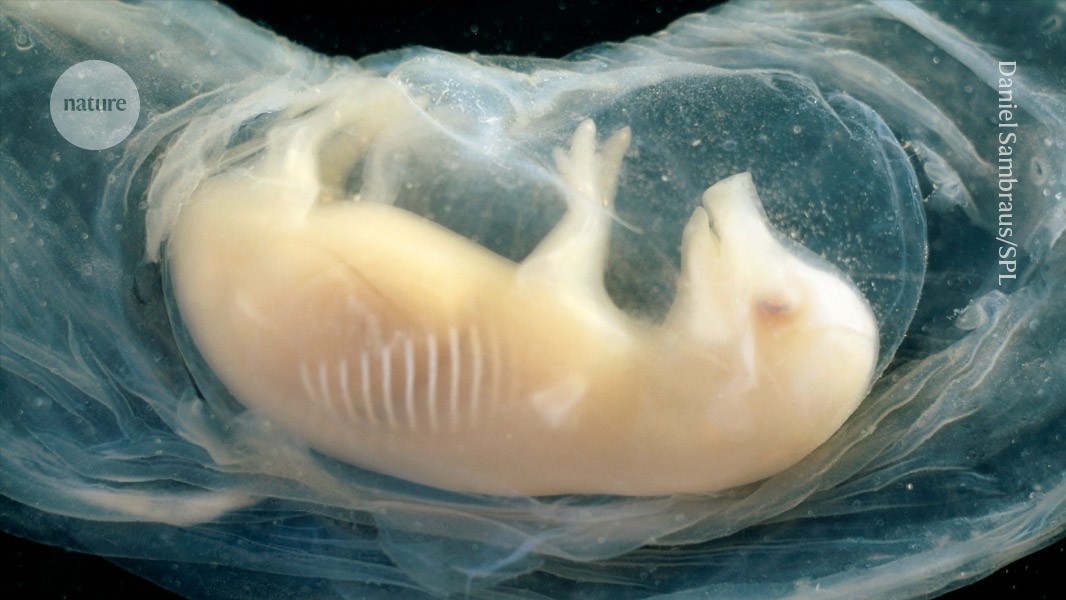
Researchers have reported growing hearts containing human cells in pig embryos for the first time. The embryos survived for 21 days, and in that time their tiny hearts started beating. The findings were presented this week at the annual meeting of the International Society for Stem Cell Research in Hong Kong.
[…]
Pigs are a suitable donor species because the size and anatomy of their organs are comparable with those of humans, says Lai Liangxue
[…]
In their study, which has not been peer reviewed, Lai and his team reprogrammed human stem cells to bolster their ability to survive in a pig, by introducing genes that prevent cell death and enhance cell growth. They then generated pig embryos in which two specific genes that have key roles in heart development were knocked out. A handful of human stem cells were introduced into the pig embryos at the morula stage, soon after fertilization — a point at which the embryo consists of a ball of about a dozen cells that are rapidly dividing. The embryos were then transferred to surrogate pigs.
The team found that the embryos grew for up to 21 days, after which they did not survive. Lai says it’s possible the human cells disrupted the function of the pig hearts.